

-
 Redaction
MIT laser reveals how drugs enter the brain
A new MIT laser self-organizes into a pencil beam, imaging the blood-brain barrier 25x faster and showing in real time how drugs enter individual brain cells.
What is the blood-brain barrier - and why is it so hard to cross?
The blood-brain barrier (BBB) is a tightly regulated physiological wall that separates the bloodstream from the brain's extracellular fluid. Its primary role is protective: it blocks pathogens, toxins, and many foreign molecules from entering the central nervous system. But this same selectivity has made it one of the most persistent obstacles in modern medicine.
The numbers here are worth sitting with for a moment, because they explain why this new MIT work matters so much. Roughly 98 percent of small-molecule drugs, and nearly all large-molecule biologics, simply cannot cross the barrier in meaningful amounts. That single fact ripples through everything downstream - it is a large part of why CNS drug candidates carry an approval rate of around 6 percent, well below the roughly 13 percent seen for drugs targeting the rest of the body. Alzheimer's research alone has absorbed tens of billions of dollars in R&D since the mid-1990s, with a failure rate north of 99 percent across that period.
So when we talk about "the blood-brain barrier problem," we are not talking about a minor formulation hurdle. We are talking about the single biggest reason that so many promising molecules for Alzheimer's disease, ALS, brain tumors, and Parkinson's disease never make it out of the lab and into a patient's bloodstream in a form the brain can actually use.
For decades, scientists have had to study this barrier after the fact, relying on end-point tissue analysis or low-resolution imaging that offered no real-time picture of how individual molecules interact with the BBB at the cellular level. You would dose a model, wait, then slice the tissue open and count what got through - a bit like trying to understand a river by only ever looking at where the water ends up, never at how it flows. A new development from MIT may be about to change that entirely.

Advanced visualization of pharmacological pathways
On April 27, 2026, a research team at the Massachusetts Institute of Technology published findings in Nature Methods that effectively allow scientists to watch the journey of drugs into the brain as it happens. The paper, led by senior author Sixian You along with first author Honghao Cao and collaborators including Roger Kamm, describes an imaging method that does not just improve on existing tools incrementally - it changes the basic question researchers are able to ask.
Until now, observing how a molecule successfully navigates - or fails to penetrate - this barrier required destructive tissue sampling or low-resolution imaging that missed the micro-scale dynamics of cellular absorption. The MIT system resolves this long-standing limitation with a counterintuitive solution rooted in nonlinear optical physics.

How the pencil beam self-organizes: the optical physics explained
The new imaging system is built around a discovery that, by the researchers' own account, ran against everything they expected to see. The story behind it is almost as interesting as the result itself. The team had already built a precise fiber shaper capable of controlling how laser light travels through a multimode optical fiber - the kind of fiber that can tolerate high power loads. While testing the limits of that fiber, lead author Honghao Cao began raising the laser power step by step, simply to see how much the setup could withstand.
The conventional wisdom in optics says that cranking up the power in this type of fiber should make the light more chaotic, not less. Instead, something unexpected happened: as the power approached a critical threshold, the scattered, disordered light suddenly snapped into a single, needle-sharp beam.
"The common belief in the field is that if you crank up the power in this type of laser, the light will inevitably become chaotic. But we proved that this is not the case. We followed the evidence, embraced the uncertainty, and found a way to let the light organize itself into a novel solution for bioimaging."
- Sixian You, assistant professor, MIT Department of Electrical Engineering and Computer Science
By directing the high-power laser into the fiber at a precise, zero-degree angle - and raising the power until the light begins to interact nonlinearly with the glass of the fiber itself - the team found that nonlinear optical effects counteract the fiber's intrinsic disorder rather than amplifying it. The result is a stable, self-localized "pencil beam" that forms without any need for custom beam-shaping components, external optics, or specialized expertise to set up.
When the researchers characterized this beam against existing alternatives, it held up remarkably well. Many conventional focused beams suffer from sidelobes - faint, blurry halos of light surrounding the main beam that degrade image sharpness. The MIT pencil beam produced none of that; it stayed clean, stable, and tightly focused even under sustained use, remaining stable for roughly 12 hours of continuous operation at the power levels used for imaging, before the researchers eventually observed irreversible fiber damage at power levels around 5.5 megawatts - well beyond the operating range needed for the technique to work.
As You put it, part of the appeal is how accessible the method is: "That is the charm of this method - you could do this with a normal, optical setup and without much domain expertise."
Imaging 25 times faster - without fluorescent tags
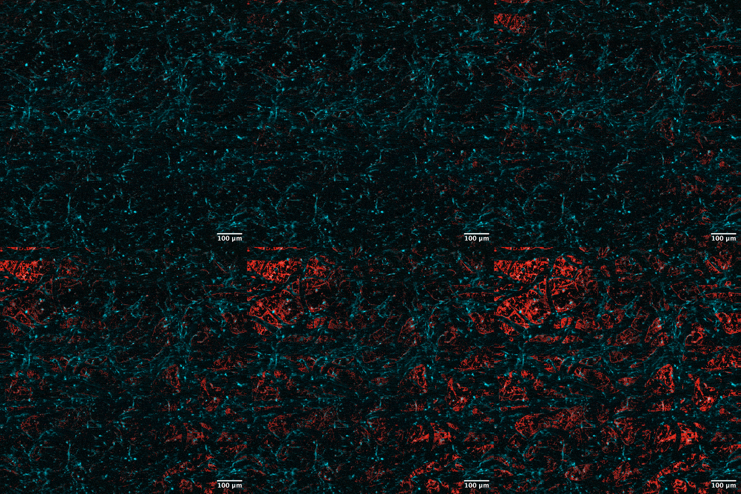
Using this self-organized pencil beam, the researchers captured three-dimensional images of a human blood-brain barrier model 25 times faster than the current gold-standard approach, while maintaining comparable image quality. That speed difference is not just a convenience - it reflects a fundamentally different way of acquiring the image.
Traditional multiphoton microscopy typically captures one thin 2D slice of tissue at a time, then requires the system to repeat that scan over and over to reconstruct a full 3D volume. It is slow, and because each slice is captured at a slightly different moment, it can blur or misrepresent processes that are actually happening dynamically, like a drug diffusing through vessel walls. The pencil beam approach instead captures volumetric, three-dimensional information from a single scan, which is what allows it to work so much faster while still resolving fine cellular detail.
Crucially, the technique does not require cells to carry fluorescent tags - enabling direct, label-free visualization of time-dependent drug entry for the first time. This matters more than it might first appear. Fluorescent labeling can itself alter the behavior of the molecule being studied, since attaching a tag changes its size, charge, and sometimes its ability to interact with cell receptors in the first place. A label-free method means researchers can watch an actual, unmodified drug candidate behave exactly as it would in the body.
The precision of this approach also allows researchers to identify which specific cell types are absorbing the pharmacological agent, including endothelial cells, pericytes, and astrocytes - the three principal cell types that together form and regulate the blood-brain barrier. Being able to distinguish uptake at this level of resolution gives an unprecedented picture of live cellular behavior, one that was previously only reconstructable indirectly, after the fact.
This marks a shift from static, post-hoc analysis to a fully kinetic model of drug absorption - one that unfolds in real time, on human-derived tissue.
Who led the research
The findings were led by Sixian You, assistant professor in MIT's Department of Electrical Engineering and Computer Science and a member of the Research Laboratory for Electronics, together with Roger Kamm, the Cecil and Ida Green Distinguished Professor of Biological and Mechanical Engineering at MIT. First author Honghao Cao carried out the foundational power-scaling experiments that led to the discovery of the pencil-beam effect, alongside co-authors Sarah Spitz, Li-Yu Yu, Kunzan Liu, Zhengyu Zhang, Federico Presutti, Francesca Michela Pramotton, and Subhash Kulkarni. Their collaboration - bridging optical physics and biomedical engineering - reflects the cross-disciplinary nature of the breakthrough, and was supported in part by MIT startup funds, the National Science Foundation, the Silicon Valley Community Foundation, the Diacomp Foundation, the Harvard Digestive Disease Core, a MathWorks Fellowship, and the Claude E. Shannon Award.
The research was published in Nature Methods and has since drawn significant attention from both the pharmaceutical development community and academic neuroscience.
Implications for drug development and delivery
In the context of the scientific discourse surrounding this publication, the development is being viewed as a transformative utility for pharmaceutical engineering. Standard drug development has historically relied on end-point analysis: researchers look at brain tissue after the fact to determine how much of a drug made it through the barrier.
The MIT system shifts this to a kinetic model, providing immediate, real-time feedback on the performance of different delivery vehicles - such as lipid nanoparticles or specialized peptide carriers - to determine which are most efficient at bypassing the BBB. By providing this feedback on human-derived tissue models, which more reliably predict clinical outcomes than animal surrogates, the iterative cycle of designing and testing new neuro-therapeutics can be shortened by months or even years.
This kind of tool is arriving at a genuinely pivotal moment for the field. Earlier in 2026, the FDA granted accelerated approval to the first biologic engineered specifically to cross the blood-brain barrier via receptor-mediated transcytosis, for a rare pediatric neurological condition. Within days, one of the world's largest pharmaceutical companies committed billions of dollars to acquire a company built around a CNS drug delivery asset. Taken together, these developments suggest that after decades of near-total failure, the barrier is no longer considered impassable - it is now considered a formulation science and imaging challenge, and that is precisely the kind of challenge a tool like the MIT pencil beam is built to help solve. If a drug candidate can be engineered to slip past the barrier, researchers now need a fast, reliable way to confirm that it actually does - and to watch, cell by cell, how it happens.
This positions the technology as a potential accelerant across nearly every stage of CNS drug development, from early-stage compound screening to preclinical delivery optimization. For readers who want a deeper foundation in how this barrier actually functions day to day - not just when something goes wrong - it's worth exploring the blood-brain barrier as a living gatekeeper, which lays out the cellular architecture behind the wall this new laser technique is now able to watch in motion.
Impact on neurodegenerative disease research
The application of this laser technology extends directly into the study of Alzheimer's disease, ALS (amyotrophic lateral sclerosis), and other neurological conditions. In these diseases, the blood-brain barrier is often compromised - or becomes hyper-selective in ways that remain poorly understood. Dementia remains, by the World Health Organization's own accounting, among the leading causes of death worldwide, and Alzheimer's disease is thought to account for the large majority of those cases. Against that backdrop, any tool that can meaningfully shorten the feedback loop between "we designed a new delivery vehicle" and "we know whether it works" carries real weight.
Discussions among the research community have highlighted how this tool enables the observation of drug efficacy in diseased versus healthy brain environments simultaneously, using human-derived in vitro models. If a researcher can observe in real time that a drug is being blocked by a specific cellular response, they can adjust the chemical composition of the treatment to mitigate that rejection.
This level of clinical precision - observing a drug's failure at the cellular level and correcting for it immediately - is what has historically been missing from the bridge between laboratory chemistry and neurological application.
Engineering challenges and the road to commercialization
While the current results are promising, the MIT team emphasized the engineering rigor required to satisfy the precise conditions that produce the pencil beam - including exact on-axis laser alignment and careful management of near-critical power levels. This is not a trivial caveat: it means the technique, while conceptually elegant, still demands a well-calibrated optical setup to reproduce reliably, and getting it wrong risks the kind of irreversible fiber damage the team observed at the upper end of the power range they tested.
As the technology is refined, the research team plans to pursue three parallel tracks:
- A deeper investigation into the fundamental physics behind pencil-beam self-organization
- Extension of the technique to imaging neurons directly in the brain, moving beyond in vitro models
- Active efforts to commercialize the technology for integration into standard laboratory workflows
This would enable researchers to observe not just the initial entry of a drug, but how its concentration and cellular impact evolve over time - providing a comprehensive pharmacokinetic profile of a medication's metabolic life cycle within the brain.
What this means for patients and the future of neuroscience
While this technology is still at the research and preclinical stage, its long-term implications for patients with neurological conditions are substantial. The primary bottleneck in developing treatments for diseases like Alzheimer's, Parkinson's, and ALS has never really been a shortage of candidate molecules - laboratories generate promising compounds constantly. The bottleneck has been the inability to efficiently determine how and whether those molecules reach the brain, and to do so quickly enough to iterate on a design before years and millions of dollars have already been spent.
By giving researchers a high-speed, high-resolution, real-time window into drug delivery dynamics at the cellular level, the MIT pencil beam system addresses this bottleneck directly. If commercialized and adopted broadly, it could meaningfully compress the timeline from laboratory discovery to clinical trial - and ultimately, to patient access.
The technology also raises broader questions about how optical physics and biomedical engineering can continue to intersect. The fact that a mechanism as fundamental as nonlinear light self-organization in an optical fiber can be repurposed into a tool for imaging human tissue at subcellular resolution is a reminder that some of the most important scientific advances emerge from the unexpected edges between disciplines - in this case, from a researcher simply turning up the power on a laser to see what would happen next.
Share now









Key takeaways
- MIT researchers discovered that chaotic laser light can spontaneously self-organize into a focused "pencil beam" inside a standard multimode optical fiber under precise power and alignment conditions - without the need for custom beam-shaping components.
- The discovery began by accident: while stress-testing a fiber's power limits, the team found that light became more ordered, not more chaotic, as power approached a critical threshold near the fiber's damage point.
- Using this self-organized pencil beam, the team captured 3D images of a human blood-brain barrier model 25 times faster than the current gold-standard approach, while maintaining comparable resolution.
- Unlike many conventional focused beams, the pencil beam produces virtually no sidelobes - the blurry halos that typically reduce image sharpness - and remained stable for roughly 12 hours of continuous operation.
- The technology enables real-time, label-free observation of individual cells absorbing drugs in human-derived in vitro BBB models - without requiring fluorescent cell tags.
- Researchers can now identify the rate at which specific cell types - including endothelial cells, pericytes, and astrocytes - internalize a pharmacological agent, providing unprecedented cellular-level detail.
- Roughly 98% of small-molecule drugs, and nearly all biologics, fail to cross the blood-brain barrier in clinically useful amounts, making CNS drug development one of pharma's toughest challenges.
- CNS drug candidates have historically carried an approval rate of around 6%, compared to roughly 13% for non-CNS drugs.
- The technique is expected to accelerate drug development for neurodegenerative diseases such as Alzheimer's and ALS by providing real-time, human-relevant feedback on drug delivery performance.
- The system exploits a nonlinear optical self-organization mechanism that transforms an ordinarily scattered, disordered laser signal into a coherent, ultrafast pencil beam capable of volumetric multiphoton imaging.
- The findings were published in Nature Methods on April 27, 2026, led by senior authors Sixian You (assistant professor, MIT EECS) and Roger Kamm (Cecil and Ida Green Distinguished Professor of Biological and Mechanical Engineering at MIT), with Honghao Cao as first author.
- Future development plans include extending the technique to direct neuron imaging in the brain and pursuing commercial integration into standard laboratory workflows.
Sources
- MIT News https://news.mit.edu/2026/self-organizing-pencil-beam-laser-could-help-scientists-design-brain-targeted-therapies-0427
- Nature Methods (original paper) https://www.nature.com/articles/s41592-026-03067-0
- ScienceDaily https://www.sciencedaily.com/releases/2026/04/260428045542.htm
- SciTechDaily https://scitechdaily.com/mit-laser-breakthrough-lets-scientists-watch-drugs-enter-the-brain-in-real-time/
- EurekAlert! https://www.eurekalert.org/news-releases/1125586
- Published 2026-05-02 15:28
- Modified 2026-08-01 20:45

-
Redaction