

-
 Redaction
Synaptic density and the aging neocortex explained
Beyond the hippocampus, the neocortex ages in complex, region-specific ways. Discover the science of synaptic density, protein turnover, and plasticity.
The conversation surrounding cognitive health tends to focus disproportionately on the hippocampus. That focus is understandable - it is, after all, the structure most closely tied to memory formation - but it leaves out most of the story. The neocortex, the vast folded sheet responsible for higher-order reasoning, sensory perception, and the traits we'd call "personality," undergoes its own distinct and rather sophisticated aging process. Maintaining synaptic density across this landscape isn't simply a matter of preventing cell death. It's a dynamic challenge involving protein turnover, structural compensation, and molecular signaling, and it deserves more attention than it typically gets in the popular conversation about brain health.
Recent clinical data has quantified this transition with what I'd call sobering precision. Research has identified a significant inverse correlation between presynaptic terminal counts and chronological age, with a correlation coefficient of r = -0.7 (p < 0.001). In plain terms: as we age, the loss of these communication hubs is predictable enough to model. By the time a person passes 60, they typically show an average 20% decrease in presynaptic terminal density compared to younger adults.
But - and this is a "but" I find myself returning to again and again in this field - that loss is not uniform. The brain isn't a monolithic structure quietly eroding at a single rate. It's more like a mosaic, made up of regions with wildly different degrees of resilience and vulnerability. Understanding where the vulnerability concentrates, and why, is where the real clinical value lies.
Quantitative loss and the sex-based neural landscape
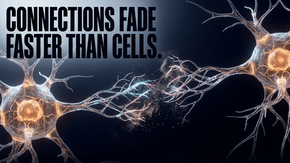
When we tally the total neural inventory, the data suggests that roughly 10% of neocortical neurons are lost across the human life span. This decline appears relatively consistent between sexes, though the starting points differ - studies have found higher total synapse and neuron counts in males compared to females, even as the rate of decline remains a shared biological reality. Worth noting: this 10% neuron loss is a broad, long-term estimate drawn from stereological studies, and figures vary depending on region and methodology. I mention this not to undermine the finding but because precision matters more than false certainty in this field.
What's more striking to me, clinically, is that this modest neuron loss often precedes or runs alongside a much larger reduction in synaptic density - the 20% figure I mentioned above. The connections between cells, in other words, tend to be more fragile than the cells themselves.
Microscopic work gives us a closer look at the texture of this decline. In the context of normal aging, researchers observe focal areas of synapse loss alongside what are called distended synaptophysin-containing boutons - swollen, less efficient versions of the structures that normally release neurotransmitter. Interestingly, even in the presence of diffuse amyloid plaques (a hallmark more associated with disease than normal aging), synaptic content often remains stable both inside and outside these plaque regions. That's a reassuring finding, in its way: it suggests the brain can maintain a baseline of synaptic health even when early histological changes are already visible. The real clinical concern lies in preventing the transition from these manageable, background changes into the more aggressive pruning that characterizes cognitive decline.
The discordance between volume loss and synaptic density
One of the more counterintuitive findings to emerge from recent neuroimaging and proteomic work is the lack of a tight correlation between brain volume and synaptic density. Take the prefrontal cortex, the seat of executive function. It experiences an age-related volume decline of roughly 4.9% per decade. To a casual observer, that number alone might suggest a fairly aggressive loss of function.
It doesn't, necessarily. When researchers measure SV2A density - a reliable proxy for synaptic vesicle protein 2A, and increasingly the gold-standard marker in PET imaging studies of synaptic health - the prefrontal cortex actually shows one of the smallest reduction rates in synaptic density among all neocortical regions.
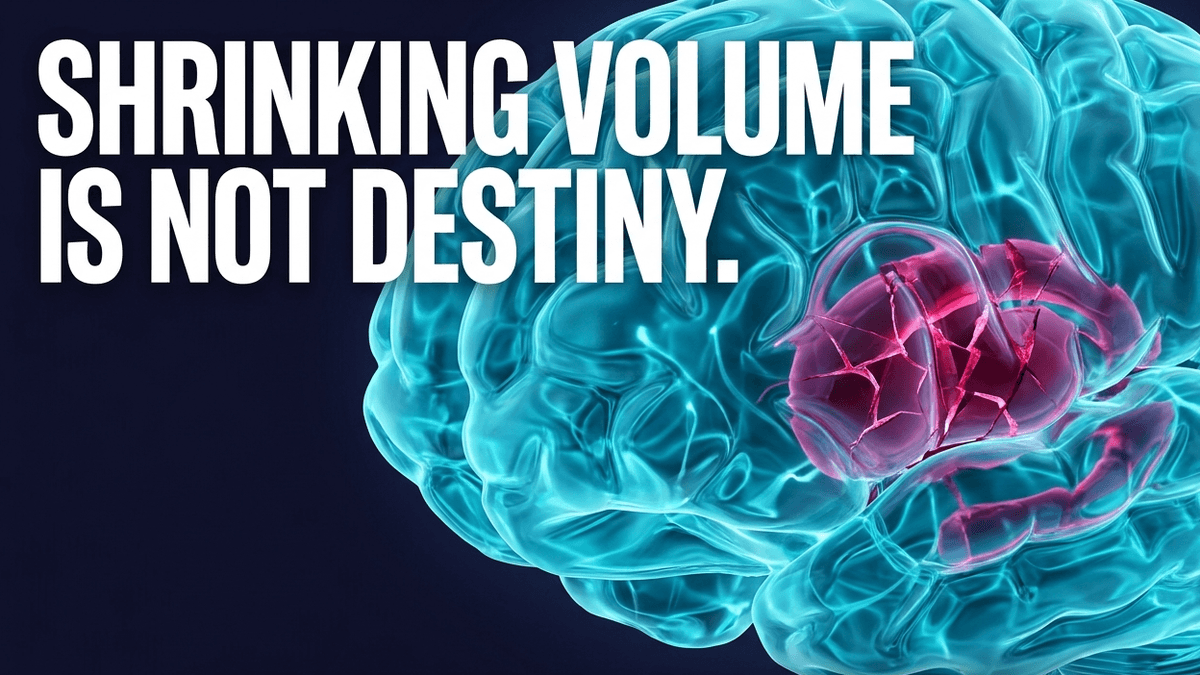
This volume-versus-density discordance suggests that the aging prefrontal cortex may be shrinking for reasons that have relatively little to do with a catastrophic loss of synaptic connections - changes in white matter integrity, water content, or glial density are more plausible culprits. Other regions tell a different story. The caudate, for instance, shows a synaptic density loss of roughly 3.6% per decade, with the medial occipital cortex following closely at 3.4%. What this tells us clinically is that a "one-size-fits-all" approach to cognitive maintenance doesn't reflect the underlying biology. Different regions age differently, and strategies for preserving them should be just as varied.
As one systematic review of in vivo SV2A PET imaging put it, the growing body of research has revealed "different spatial patterns of synaptic density loss" across neurodegenerative conditions - a finding that extends, in more modest form, to healthy aging itself.
Regional vulnerability and the brain's compensation mechanism
The brain does not passively accept the loss of its connections. It engages in what clinicians describe as compensatory responses - a kind of internal negotiation between decline and adaptation. In the precentral cortex, for example, evidence suggests that synaptic plasticity within the synaptic contact zone acts as a direct countermeasure to regional synapse loss. When one connection weakens, nearby structures may reorganize or strengthen to pick up the slack. This is, in essence, the biological substrate of what we call cognitive reserve.
Not every region is equally adaptable, though. The somatosensory cortex - responsible for touch and bodily position sense - shows virtually no age-related synapse loss at all. Because this region stays so stable, it shows comparatively little visible plasticity; it simply doesn't need to compensate for damage that isn't occurring.
The dorsolateral prefrontal cortex tells a more concerning story. Studies in aged monkeys have linked cognitive impairment specifically to the loss of "thin spines" in layer 3 of the cortex - small, motile dendritic protrusions thought to be the physical substrate of new learning. Larger, more stable spines tend to persist even as these thin spines disappear. It's the loss of flexibility, not just volume, that appears to drive age-related forgetfulness in this region. I find this distinction clinically important: it reframes the goal of cognitive maintenance away from simply "preserving brain tissue" and toward preserving the kind of tissue that supports ongoing learning.
The molecular burden of slowed protein turnover
To understand why synapses fail, you have to look at the protein level. Aging is fundamentally associated with a reduction in the breakdown - and thus the efficient recycling - of synaptic proteins. In a younger brain, proteins are built, used, and cleared with impressive efficiency. In the aging neocortex, this turnover slows considerably.
The consequence is that long-lasting, aged proteins are more likely to aggregate, or to "spill over" and be taken up by microglia, the brain's resident immune cells. When microglia encounter this aggregated material in excess, they can become overactive - triggering a dysfunctional, overly aggressive pruning of synapses that are otherwise perfectly healthy. It's a case of the clean-up crew getting overzealous.

This proteomic shift carries several molecular hallmarks:
- An age-dependent decrease in the expression of synaptophysin, a core presynaptic vesicle protein
- Reduced postsynaptic density protein 95 (PSD-95), critical for organizing receptors at the synapse
- Declines in the SNARE family proteins, the molecular machinery that fuses vesicles to the cell membrane for neurotransmitter release
When these components decline, a neuron's ability to release and receive neurotransmitters is measurably compromised. Across both human tissue studies and experimental animal models, dendritic spine reductions in the cortex and hippocampus range from roughly 20% to 40% during normal aging - a wide range, reflecting real variability across studies, regions, and measurement techniques, but a consistent signal nonetheless.
Vesicle-cycle modeling and the demand for new vesicles
Recent mathematical models have shed useful light on the activity-dependent aging of synaptic vesicles. I like to think of a synaptic vesicle as a delivery truck. In a highly active synapse, these trucks are on the road constantly. Over time, the proteins that make up the truck itself begin to wear down. If the cell can't supply freshly built vesicles quickly enough to replace the aging fleet, signal transmission becomes sluggish, or fails outright.
This demand-dependent supply chain is a critical, underappreciated factor in maintaining robust neural transmission. It also explains why high cognitive activity is something of a double-edged sword: it keeps the system in active use, which is generally good, but it also raises the demand for protein replacement. If the biological cleanup and rebuilding crew isn't keeping pace - due to poor sleep, metabolic strain, or other stressors - the system can begin to fail under its own workload.
This is where molecular regulators like the p75 neurotrophin receptor (p75NTR) enter the picture. p75NTR acts as a negative regulator of structural and functional plasticity, and its expression rises measurably with age. In studies of mutant mice lacking p75NTR, researchers found the animals were resistant to several age-associated disruptions of plasticity in the hippocampus - including deficits in long-term potentiation and associative memory. That work has so far centered on the hippocampus rather than the neocortex broadly, so I'd stop short of generalizing the finding outward. Still, it's a compelling proof of concept: by modulating pathways like BDNF, MAPK, and Arc, we may eventually find pharmacological ways to unlock more of the brain's youthful capacity to rewire itself, in the cortex as well as in memory-related structures.
Strategies for maintenance: the role of BDNF and CSF flow
While editing our own receptors at home remains firmly in the realm of science fiction, we've identified several powerful behavioral levers that stimulate the generation of Brain-Derived Neurotrophic Factor (BDNF) - and exercise is perhaps the most well-documented of these.
Physical activity increases BDNF expression not just in the hippocampus, where its effects are most studied, but within the broader cortex as well. BDNF acts something like fertilizer for neural tissue, encouraging the growth and maintenance of the very dendritic spines that aging threatens. Human studies in older adults have shown that structured aerobic programs - walking at a moderate pace several times a week is a well-studied example - can meaningfully raise circulating BDNF and support functional brain changes. The dose-response relationship isn't fully mapped, and results vary by intensity, frequency, and individual metabolic factors, but the general direction of the evidence is consistent: movement matters for the aging cortex, not just the aging hippocampus.
The second major lever is the biochemical cleaning that happens during sleep. Cerebrospinal fluid movement through the brain's tissue increases significantly during deep, slow-wave sleep, as part of what researchers call the glymphatic system - a network first characterized in 2012 that uses perivascular channels to flush metabolic waste out of brain tissue. During this stage, the space between neurons expands by an estimated 60%, dramatically lowering the resistance to fluid flow and allowing waste, including the aged, aggregated proteins discussed earlier, to be cleared more efficiently. That waste is ultimately collected by the meningeal lymphatic vessels and carried toward the deep cervical lymph nodes, near the base of the neck, rather than draining directly into a single vessel.
Without adequate slow-wave sleep, the neocortex is left to simmer for longer in its own metabolic byproducts - and slow-wave sleep is, unfortunately, one of the sleep stages that declines most steeply with age. This creates something of a vicious cycle: aging reduces the very sleep architecture the brain needs to clear the debris that aging itself produces. It's one of the more compelling arguments I've encountered for treating sleep quality as a genuine clinical priority in cognitive aging, rather than a lifestyle afterthought.

Novelty, bilingualism, and the engagement of multiple networks
Finally, the maintenance of synaptic density is heavily influenced by how we use our brains day to day. Novelty and complex social information processing are two of the most reliable ways to simultaneously engage multiple cortical networks at once. Learning a new language, navigating an unfamiliar city, or working through a genuinely complex social interaction doesn't just recruit one brain region - it demands coordinated effort across large swaths of the neocortex.
Bilingualism offers a particularly well-studied example of this principle in action. Longitudinal and cross-sectional research has linked sustained bilingual language use to greater preservation of cortical thickness in aging, along with a delay in the onset of dementia symptoms estimated at several years in some cohort studies. The proposed mechanism involves ongoing synaptic reorganization - new connections forming, useful ones being reinforced, and inefficient ones being pruned - a process that closely mirrors the broader "use it or lose it" principle governing cortical maintenance throughout life.
This multi-network engagement is, in effect, a biological imperative. Plasticity is at its most vigorous during development, but the data is fairly clear that it persists throughout the lifespan, continuing to reorganize structure and function well into older age. The aging neocortex isn't a passively shrinking organ resigned to decline. It's a dynamic structure that, given the right metabolic, physical, and cognitive inputs, retains a genuine capacity to maintain the connections that make us who we are.
Bringing it together
The picture that emerges from this research isn't one of inevitable, uniform decline - it's one of regional variability, molecular nuance, and real opportunity for intervention. Protein turnover, sleep architecture, and BDNF stimulation through movement and mental engagement aren't just wellness buzzwords; they map directly onto the mechanisms this research has identified. Moving beyond the superficiality of brain games and toward a genuine, biologically grounded understanding of how the neocortex ages is, I think, the more honest and ultimately more useful conversation to be having.
Share now









Key takeaways
- NIH/PMC - https://pmc.ncbi.nlm.nih.gov/articles/PMC7884039/
- Journal of Nuclear Medicine (SNMMI) - https://jnm.snmjournals.org/content/66/4/620
- Frontiers in Aging Neuroscience - https://www.frontiersin.org/journals/aging-neuroscience/articles/10.3389/fnagi.2024.1428244/full
- NIH/PMC - https://pmc.ncbi.nlm.nih.gov/articles/PMC10940592/
- PNAS - https://www.pnas.org/doi/10.1073/pnas.1610909114
Sources
- usf.edu https://digitalcommons.usf.edu/cgi/viewcontent.cgi?article=4812&context=ujmm
- nih.gov https://pubmed.ncbi.nlm.nih.gov/8423884/
- nih.gov https://pubmed.ncbi.nlm.nih.gov/9215725/
- nih.gov https://pmc.ncbi.nlm.nih.gov/articles/PMC12701775/
- semanticscholar.org https://www.semanticscholar.org/paper/Synaptic-density-in-human-frontal-cortex-changes-of-Huttenlocher/d753094372f1b27015c7109ccfc265e4815e405d
- nih.gov https://pmc.ncbi.nlm.nih.gov/articles/PMC3592200/
- nih.gov https://pmc.ncbi.nlm.nih.gov/articles/PMC10940592/
- nih.gov https://pmc.ncbi.nlm.nih.gov/articles/PMC11330810/
- frontiersin.org https://www.frontiersin.org/journals/aging-neuroscience/articles/10.3389/fnagi.2024.1428244/full
- mbexc.de https://mbexc.de/how-synapses-maintain-their-activity-despite-ageing-2/
- nih.gov https://pmc.ncbi.nlm.nih.gov/articles/PMC7884039/
- Published 2026-07-30 14:19
- Modified 2026-07-30 14:19

-
Redaction